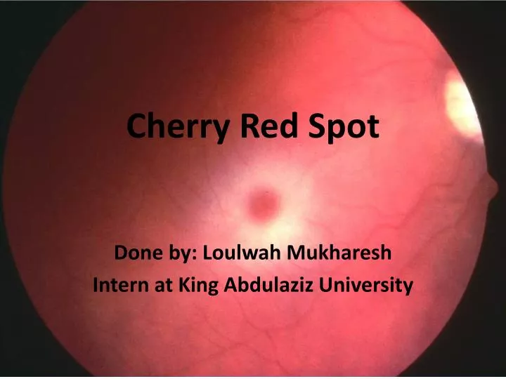
Cherry Red Spot / Mancha rojo cereza - Cherry-red spot - qaz.wiki / They're nothing to worry about from a medical.
Cherry Red Spot / Mancha rojo cereza - Cherry-red spot - qaz.wiki / They're nothing to worry about from a medical.. The appearance of a cherry red spot at the macula, caused by the contrast of a red macula against retinal pallor, occurs in a number of metabolic storage disorders, including: These red spot lesions can occur at any part of the body they may appear in different sizes and can be single at one location or multiple and cherry angioma. Sialidosis (type i = cherry. It occurs in nearly 100% of patients with type i while only 75% of type ii patients have this feature possibly because their early. Loulwah mukharesh intern at king abdulaziz university.
Branch retinal artery occlusion (brao). 15.58) is a clinical sign seen in the context of thickening and loss of transparency of the retina at the posterior pole. Loulwah mukharesh intern at king abdulaziz university. Caused by swelling of retinal nerve fibers due to ischemia. The appearance of a cherry red spot at the macula, caused by the contrast of a red macula against retinal pallor, occurs in a number of metabolic storage disorders, including:
Cherry red spot in CRAO - American Academy of Ophthalmology from www.aao.org They're nothing to worry about from a medical. They range from the size of a pinpoint up to ¼ of an inch in diameter. It occurs in nearly 100% of patients with type i while only 75% of type ii patients have this feature possibly because their early. These are tine, red moles on the skin. Call them cherry angiomas, red freckles, or red moles, these red spots are an annoying—and unsightly—condition to deal with. A cherry red spot is may be seen in late childhood or early adolescence. Although they can appear anywhere on the body, they commonly appear on the face, lip, torso, and scalp. Cherry angiomas are bright cherry red, usually oval or circular, and small.
Red moles, or cherry angiomas, are common skin growths that can develop on most areas of your they're also known as senile angiomas or campbell de morgan spots.
Residents and fellows contest rules | international ophthalmologists contest rules. Branch retinal artery occlusion (brao). Loulwah mukharesh intern at king abdulaziz university. These red spot lesions can occur at any part of the body they may appear in different sizes and can be single at one location or multiple and cherry angioma. A cherry red spot is may be seen in late childhood or early adolescence. They're nothing to worry about from a medical. Cherry angiomas are bright cherry red, usually oval or circular, and small. These are tine, red moles on the skin. 15.58) is a clinical sign seen in the context of thickening and loss of transparency of the retina at the posterior pole. Eye, startle responses in infancy, neurodegeneration, and death. Call them cherry angiomas, red freckles, or red moles, these red spots are an annoying—and unsightly—condition to deal with. There is no cherry red spot and there is only partial visual field loss. The appearance of a cherry red spot at the macula, caused by the contrast of a red macula against retinal pallor, occurs in a number of metabolic storage disorders, including:
Moreover, i've read that the red color comes from the choroid blood vessels, but isn't the choroid black? It occurs in nearly 100% of patients with type i while only 75% of type ii patients have this feature possibly because their early. They range from the size of a pinpoint up to ¼ of an inch in diameter. There is no cherry red spot and vision is reduced to light perception. They're nothing to worry about from a medical.
PPT - Cherry Red Spot PowerPoint Presentation, free ... from image1.slideserve.com Caused by swelling of retinal nerve fibers due to ischemia. Call them cherry angiomas, red freckles, or red moles, these red spots are an annoying—and unsightly—condition to deal with. Loulwah mukharesh intern at king abdulaziz university. There is no cherry red spot and vision is reduced to light perception. It describes the appearance of a small circular choroid shape as seen through the fovea centralis. Although they can appear anywhere on the body, they commonly appear on the face, lip, torso, and scalp. So what are those little red spots anyway? Branch retinal artery occlusion (brao).
Loulwah mukharesh intern at king abdulaziz university.
These are tine, red moles on the skin. So what are those little red spots anyway? So what is the explanation of a cherry red spot? 15.58) is a clinical sign seen in the context of thickening and loss of transparency of the retina at the posterior pole. Branch retinal artery occlusion (brao). Moreover, i've read that the red color comes from the choroid blood vessels, but isn't the choroid black? Residents and fellows contest rules | international ophthalmologists contest rules. Sialidosis (type i = cherry. Caused by swelling of retinal nerve fibers due to ischemia. The appearance of a cherry red spot at the macula, caused by the contrast of a red macula against retinal pallor, occurs in a number of metabolic storage disorders, including: It describes the appearance of a small circular choroid shape as seen through the fovea centralis. There is no cherry red spot and vision is reduced to light perception. Red moles, or cherry angiomas, are common skin growths that can develop on most areas of your they're also known as senile angiomas or campbell de morgan spots.
Cherry angiomas are bright cherry red, usually oval or circular, and small. So what is the explanation of a cherry red spot? Branch retinal artery occlusion (brao). The retinal opacification may be due to various causes. Eye, startle responses in infancy, neurodegeneration, and death.
Cherry-red spot in Tay-Sachs disease. The right frame ... from www.researchgate.net They're nothing to worry about from a medical. Branch retinal artery occlusion (brao). These are tine, red moles on the skin. Eye, startle responses in infancy, neurodegeneration, and death. It describes the appearance of a small circular choroid shape as seen through the fovea centralis. It occurs in nearly 100% of patients with type i while only 75% of type ii patients have this feature possibly because their early. The appearance of a cherry red spot at the macula, caused by the contrast of a red macula against retinal pallor, occurs in a number of metabolic storage disorders, including: Although they can appear anywhere on the body, they commonly appear on the face, lip, torso, and scalp.
Cherry angiomas are bright cherry red, usually oval or circular, and small.
Sialidosis (type i = cherry. 15.58) is a clinical sign seen in the context of thickening and loss of transparency of the retina at the posterior pole. It occurs in nearly 100% of patients with type i while only 75% of type ii patients have this feature possibly because their early. Residents and fellows contest rules | international ophthalmologists contest rules. Although they can appear anywhere on the body, they commonly appear on the face, lip, torso, and scalp. Caused by swelling of retinal nerve fibers due to ischemia. They range from the size of a pinpoint up to ¼ of an inch in diameter. A cherry red spot is may be seen in late childhood or early adolescence. Branch retinal artery occlusion (brao). Loulwah mukharesh intern at king abdulaziz university. Red moles, or cherry angiomas, are common skin growths that can develop on most areas of your they're also known as senile angiomas or campbell de morgan spots. The retinal opacification may be due to various causes. Moreover, i've read that the red color comes from the choroid blood vessels, but isn't the choroid black?
Although they can appear anywhere on the body, they commonly appear on the face, lip, torso, and scalp cherry. Sialidosis (type i = cherry.